
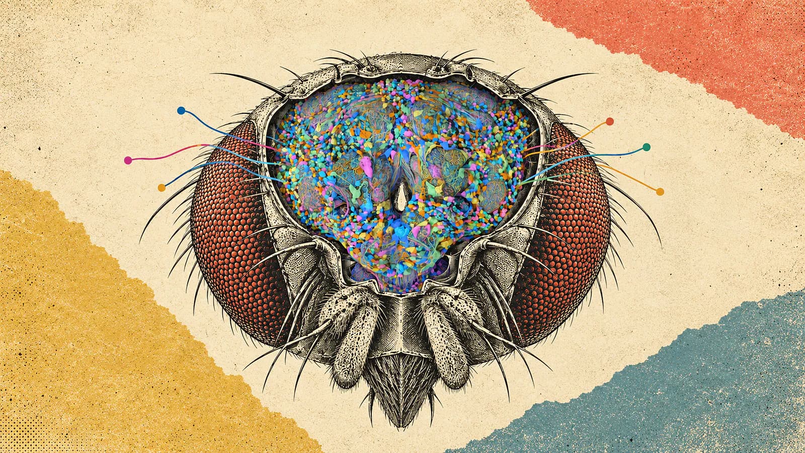
Google Research and its collaborators have mapped the complete central nervous system of an adult male fruit fly, including its brain and the nerve cord connecting it to the body.
The reconstruction contains more than 166,000 neurons and approximately 125 million synaptic connections. That’s a ridiculous amount of detail inside an animal most of us only notice when it gets near our food.
A few months ago, I wrote about researchers putting a computational fruit fly brain inside a simulated body. For context, this is what a fruit fly looks like:
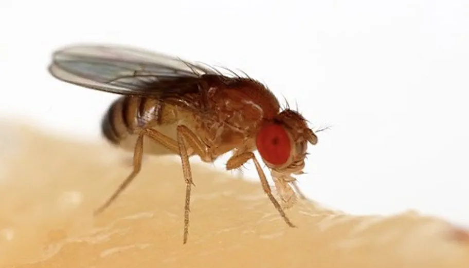
Sample image of a fruit fly
That project immediately came to mind when I saw this announcement.
We already have researchers trying to make biological wiring run in software, and now they have another complete nervous system to investigate.
The two projects tackle different problems. The simulation work explored what a model built from existing brain connections could do. Google and its partners have reconstructed the connections themselves, including pathways between the brain and the body’s movement circuits.
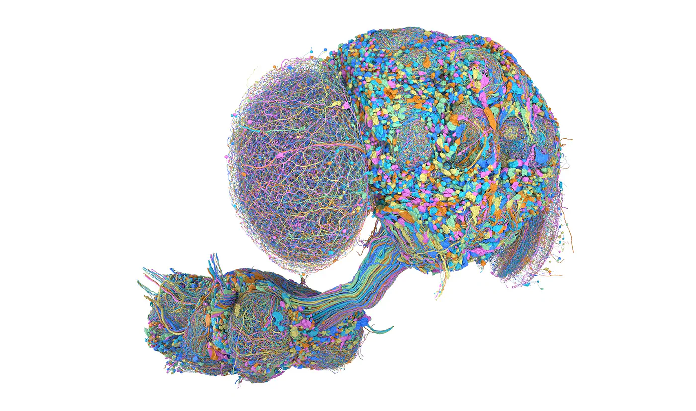
Selected neurons in the male fruit fly’s brain and ventral nerve cord, which is analogous to a spinal cord.
I’m interested in how those efforts come together. A digital fly moving around is fun to watch, but being able to explain why it moves that way would be much more useful.
What’s New About Another Fruit Fly Brain Map?
Researchers already published a complete adult female fruit fly brain map in 2024. That was the FlyWire resource behind the computational model I covered earlier, so anyone wondering whether we’ve heard this story before has a fair point.
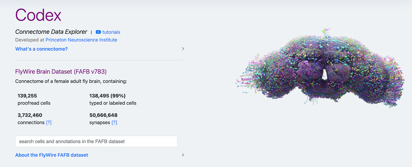
Connectome data explorer of a FlyWire brain dataset
Users on Reddit asked exactly that. In the discussion about Google’s announcement, some questioned how the work differed from previous connectomes. Others pointed out that comparing male and female brains gives scientists something a single reconstruction couldn’t provide.
This project also covers the central brain, optic lobes, and ventral nerve cord, with the connection through the neck preserved. Researchers can follow circuits across those structures within one animal.
The work was led by HHMI’s Janelia Research Campus, with contributions from Google Research, the University of Cambridge, and other collaborators. Google helped reconstruct neural structures using AI, while specialist teams checked and annotated the results. There’s a lot of human work behind those colorful images.
One thing that shouldn’t be missed is the timing. Google’s September 3 announcement accompanies publication in Cell, but earlier versions of the dataset were already available. Janelia lists an initial MaleCNS v0.9 release in October 2025, followed by v1.0 in June 2026.
So this is the formal publication and its findings getting wider attention. It also sits alongside other connectomics projects, including a recently released female brain-and-nerve-cord reconstruction. The male map adds another specimen and a new basis for comparison to that growing collection.
Why Including the Nerve Cord Is a Big Deal
A connectome is a map of neurons and the synapses connecting them. To build one, researchers trace cells through three-dimensional tissue and identify where they communicate with other cells.
The ventral nerve cord has a role broadly comparable to a spinal cord. It contains circuits involved in controlling movement, which makes it especially relevant when you want to understand how something the fly sees or senses becomes a physical action.
That’s also why the headline neuron count needs a little context. The more than 166,000 neurons cover the brain and nerve cord together.
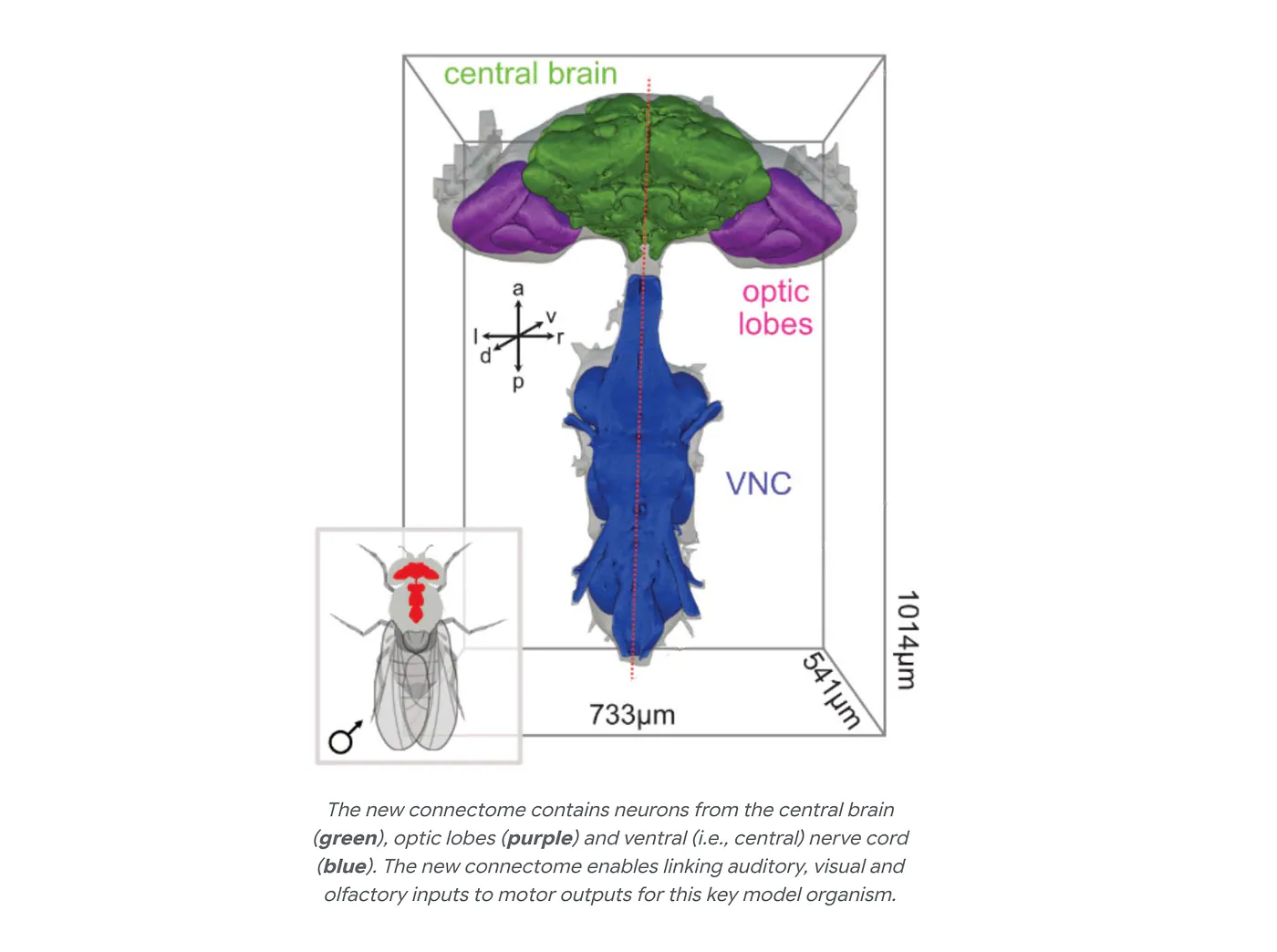
Labeled diagram showing the central brain in green, optic lobes in purple, and ventral nerve cord in blue.
Think about a fly noticing another fly nearby and turning toward it. Researchers need to follow the visual signal through the brain, identify the pathways carrying commands downward, and examine how those commands reach movement-related circuits.
A brain-only map leaves some of that investigation outside its boundaries. Having the structures connected in one specimen makes the route easier to trace.
This connects directly to the earlier simulation story. Eon Systems used selected outputs from its brain model to influence controllers for a virtual fly body. Its technical explanation says the simulation didn’t include the complete downstream motor hierarchy of a biological fly.
This is incredibly interesting.
The male reconstruction gives researchers additional anatomy to investigate when building models of how brain signals become movement. It won’t automatically replace the engineered controllers, but it provides connections against which researchers can check their assumptions.
I’d want to compare the two approaches. How much behavior can a simplified controller explain, and where does adding the mapped nerve-cord circuitry improve the prediction? That would tell us which biological details are necessary for the behavior being studied.
AI Traced the Cells, but People Had to Check Them
The reconstruction starts with electron microscopy images of physical tissue. Researchers then use computational methods to identify neural structures and assemble them into a three-dimensional reconstruction.
Google’s connectomics tools include flood-filling networks. These start from a small location inside an object and repeatedly predict which surrounding pixels belong to it. As the predicted region expands, the software traces the structure through the image volume.
The difficult part is keeping track of thin, branching neurons packed closely together. A branch has to remain assigned to the correct cell across many images, even when neighboring structures look similar.
Get that wrong and the connectivity analysis can go wrong too. Joining two separate cells creates an incorrect reconstruction; splitting one cell can make part of its structure appear disconnected.
For anyone who works with data, the problem should sound familiar. An error introduced during processing can survive into later analysis and look like a meaningful result.
Here, that could affect which cells appear to communicate.
This is where expert proofreading becomes essential. Scientists need to inspect and correct the reconstruction before relying on it to investigate circuits.
I like that the project gives this work attention. It’s easy to look at the final rendering and assume an AI processed some images and handed over a finished brain. The years of verification and annotation are a major part of making the dataset usable.
From a software perspective, I’d judge improvements by how much reliable reconstruction they produce for the human effort involved. Processing more images per hour helps, but difficult errors can consume whatever time the faster model saved.
It also affects simulations built from the data. A computational neuron model can follow its instructions perfectly while producing misleading results because the underlying wiring is wrong. Better reconstruction improves the starting material for everyone downstream.
Small Differences Can Affect Much Larger Circuits
The male–female comparison lets researchers investigate more than whether a particular neuron exists in both animals. They can examine its branches, its partners, and where information travels next.
Janelia reports 262 male-specific and 114 sexually dimorphic cell types. Dimorphic types occur in both sexes but differ in their anatomy or connectivity. These categories describe types of cells, rather than individual neuron counts.
Many of the differences appear in higher-order regions involved in behavioral control. Sensory and motor regions are largely shared, while differing connections allow relatively small neural populations to influence larger circuits.
What’s interesting is how much depends on where those cells connect. Knowing that a neuron exists tells you something. Knowing which signals reach it and which downstream cells it influences gives you a much better basis for investigating its role.
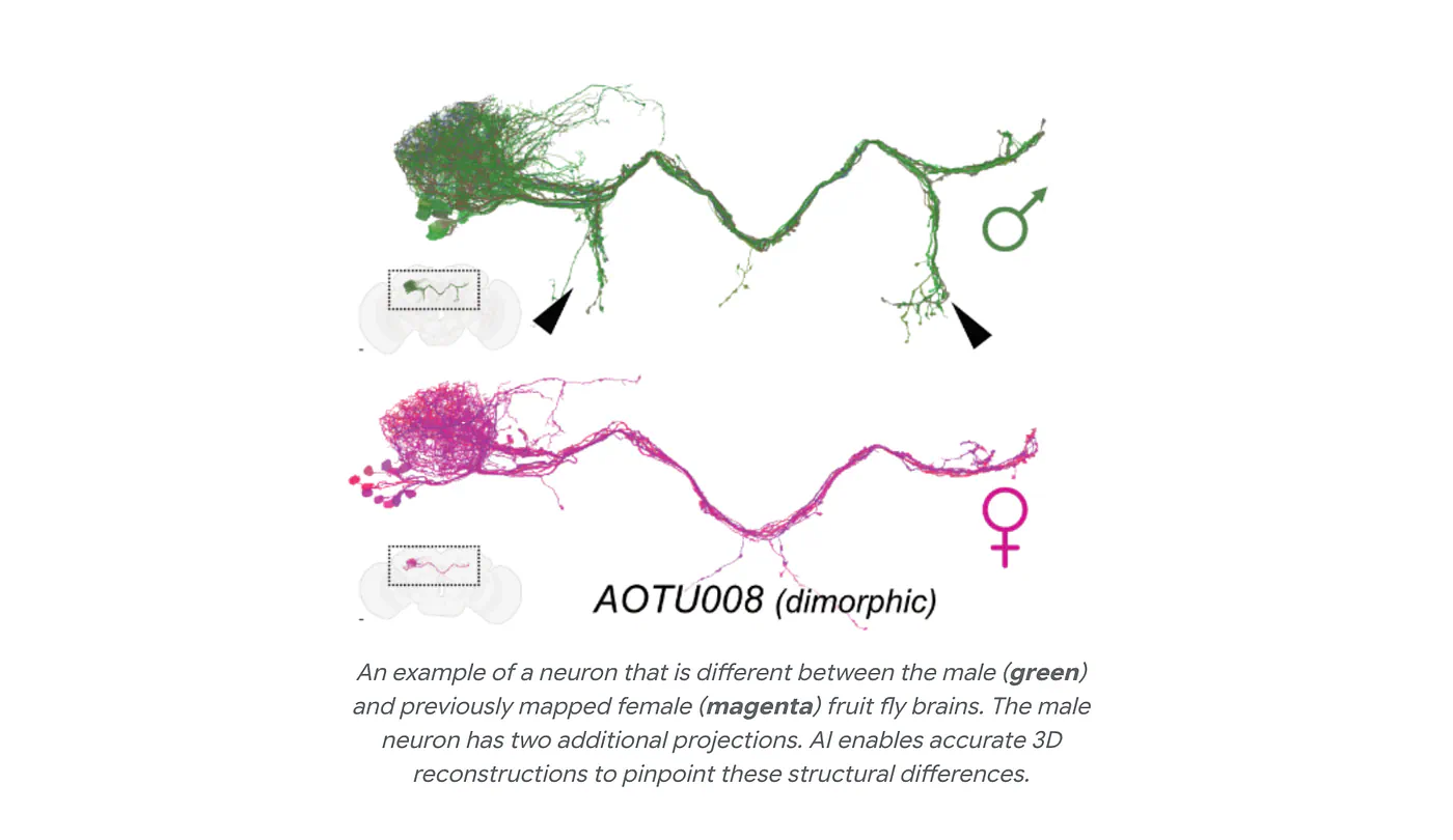
Male and female AOTU008 neurons overlaid, with the male neuron in green and female neuron in magenta.
Google’s visuals show this through an AOTU008 neuron whose male version has two additional projections. Another example, AOTU012, illustrates how neurons present in both sexes can have a mixture of shared and differing connection partners.
I’d be curious to see researchers carry those differences into computational experiments. Keep the modeling assumptions consistent, change the relevant connections, and examine whether the predicted response changes.
That would extend the kind of work done with the earlier female brain model. Researchers would have an anatomical reason to compare two versions of a circuit, followed by a specific prediction to test in living flies.
There’s still individual variation to investigate. Additional specimens would help establish which differences consistently separate the sexes and which vary between animals of the same sex.
But having a second complete reconstruction already makes that question possible to ask in much greater detail. I don’t think every connectome needs to introduce a dramatically different animal to justify the work.
The Earlier Brain Model Already Made Testable Predictions
The research I covered a few months ago helps explain why these maps are useful beyond visualization.
In the 2024 Nature paper, Philip Shiu and collaborators built a computational model using the female FlyWire connectome and predicted neurotransmitter identities. The model investigated how sensory stimulation propagated through circuits involved in feeding and grooming.
They used a leaky integrate-and-fire model. In basic terms, each simulated neuron accumulates incoming signals, loses some of that accumulated activity over time, and fires when it reaches a threshold.
It’s a simplified neuron, but the connections come from the reconstructed brain. That combination lets researchers investigate how much of a circuit’s response they can predict using its wiring and a relatively simple description of neural activity.
The sugar-sensing experiment makes this easier to understand. A real fly detects sugar through taste neurons, which send signals into circuits involved in extending its proboscis, the organ it uses to feed.
Researchers activated corresponding taste neurons in the model and examined the response downstream. The model predicted neurons involved in taste processing and feeding initiation, and the team tested predictions through biological experiments, including optogenetic activation.
That’s the part I find compelling. You can use the model to identify a candidate neuron, interfere with it experimentally, and see whether the animal responds as predicted.
The public brain-model repository supports activating and silencing selected neurons, then examining spike times and firing rates. Researchers can investigate the effects of removing particular cells from the simulated circuit before planning a biological experiment.
Drosophila Brain ModelGoogle’s new reconstruction expands the anatomical material available for this kind of work. It also adds the possibility of comparing corresponding male and female pathways.
I’d especially like to know where a model that works well on the female connectome stops predicting male responses accurately. A mismatch could help identify which connection differences deserve closer experimental attention.
About That Fly Inside a Computer
Users on X were already discussing simulated fly brains months before Google’s announcement. Eon’s demonstration helped attract that attention, showing a virtual fly navigating toward food, grooming, and beginning to eat. The company later published a technical explanation of how it worked.
The body came from NeuroMechFly and ran in the MuJoCo physics engine. Sensory events activated pathways in the brain model, selected outputs influenced movement, and the resulting movement changed the sensory input.
That feedback loop is an interesting experiment in its own right. The brain model has consequences for what happens in the simulated environment.
There’s something I’d correct from my earlier coverage, though. Describing the fly as walking, grooming, and feeding without any training overstated what the complete system demonstrated.
Eon says its body controllers were trained through imitation learning. Selected neural outputs influence those controllers, which translate the signals into movements. The brain model’s connectivity and the body’s movement machinery therefore have different origins.
I still think the demonstration is cool. I just find the actual implementation more interesting than calling it a brain that woke up inside a computer.
It gives researchers an environment for testing how a connectome-based model interacts with a body. They can change the sensory input, alter the neural model, or revise the interface and examine what happens.
The new male map could support more detailed versions of those experiments. Researchers now have connected brain-and-nerve-cord anatomy to consult when deciding how signals should pass toward movement circuits.
For me, the useful comparison would be between increasingly biological models of the same behavior. Does adding circuit detail improve the match with a living fly, or merely make the simulation more expensive? A realistic-looking animation alone can’t answer that.
You Can Explore the Data Yourself
Janelia provides tools for browsing the reconstruction and querying its connections, including neuPrint, Neuroglancer, and a cell-type explorer. Researchers can also access data programmatically and download resources for their own analyses.
The Male CNS project page is a useful starting point for finding those tools and the accompanying visualizations.
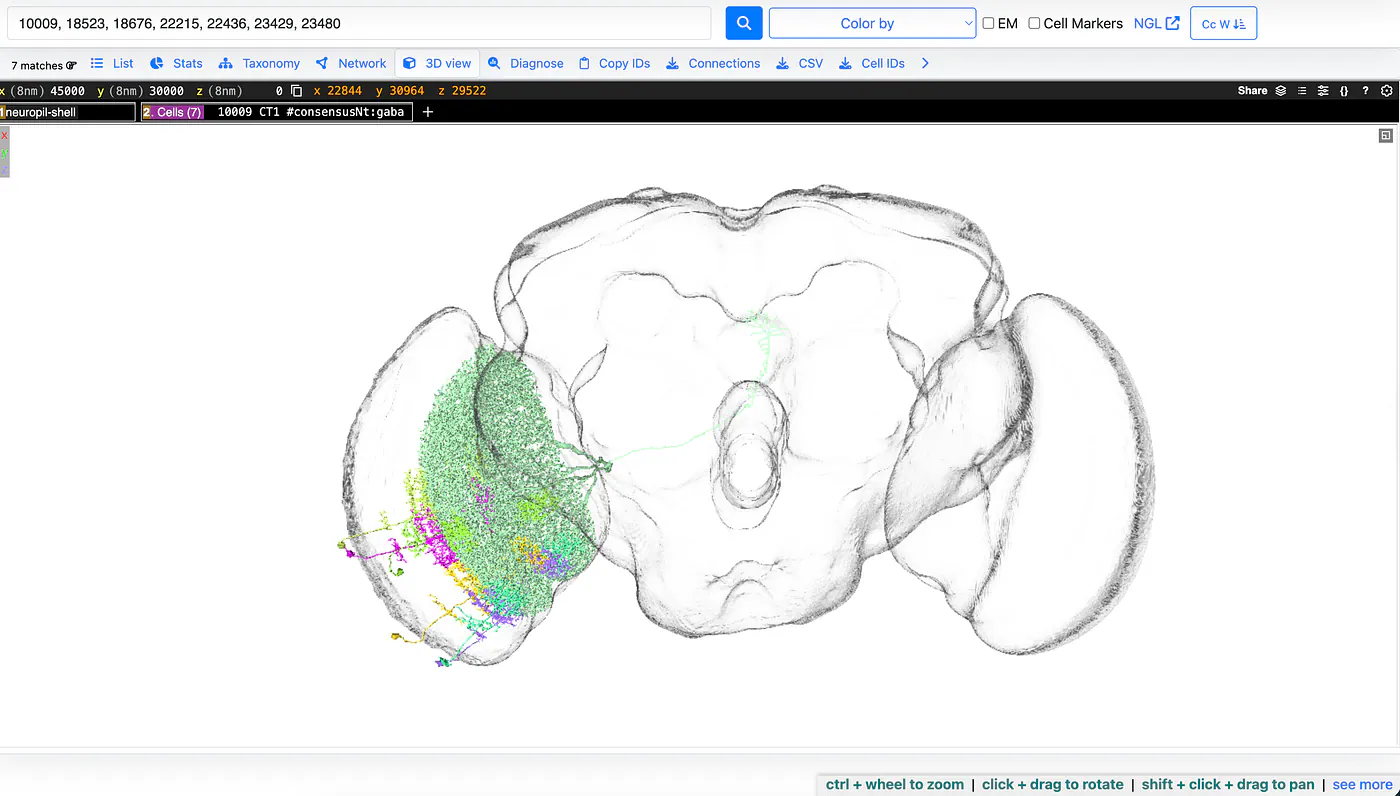
The Male CNS project simulation of a fruit fly’s brain
I’d start with one pathway rather than trying to make sense of the entire colorful reconstruction. Choose a cell associated with a sensory input or behavior, inspect its partners, and follow where those connections lead.
Google’s examples include a visual pathway involving the male-specific LoVP92 neuron, associated with courtship circuitry. That gives readers a concrete route to explore instead of an overwhelming collection of thousands of cells.
For someone coming from software, the earlier brain-model repository offers another entry point. It includes an example notebook for manipulating neural activity and examining the output.
I like having both options. You can inspect the anatomy or work with a model that uses anatomical data. Keeping track of the dataset version and the modeling assumptions would be part of any meaningful comparison between them.
Final Thoughts
I’m more interested in this announcement because I’ve already written about the simulated fly. That earlier project showed me why researchers want these maps, and the male reconstruction adds detail exactly where the relationship between a brain and a moving body becomes important.
The male–female comparison is also interesting. Researchers can investigate why similar nervous systems produce different responses and test whether particular connections help explain them.
I don’t need this to promise a human brain upload to find it exciting. Predicting what happens when a specific fly neuron is activated or silenced is already a substantial scientific result.
What I’d like to see next is a simulation that uses the mapped brain and nerve cord to predict a movement response, followed by an experiment that checks it in a living fly.
Then we can examine which parts of the model explained the behavior and which parts still need work.
What do you think? How far are we from uploading a human brain to a digital simulation?
References:
Sources
- mapped the complete central nervous system of an adult male fruit flyblog.google
- researchers putting a computational fruit fly brain inside a simulated bodyx.com
- discussion about Google’s announcementreddit.com
- connectomecell.com
- 2024 Nature papernature.com
- https://www.youtube.com/watch?v=bZVoPJumx8Y&t=4syoutube.com
- public brain-model repository


